Meaning
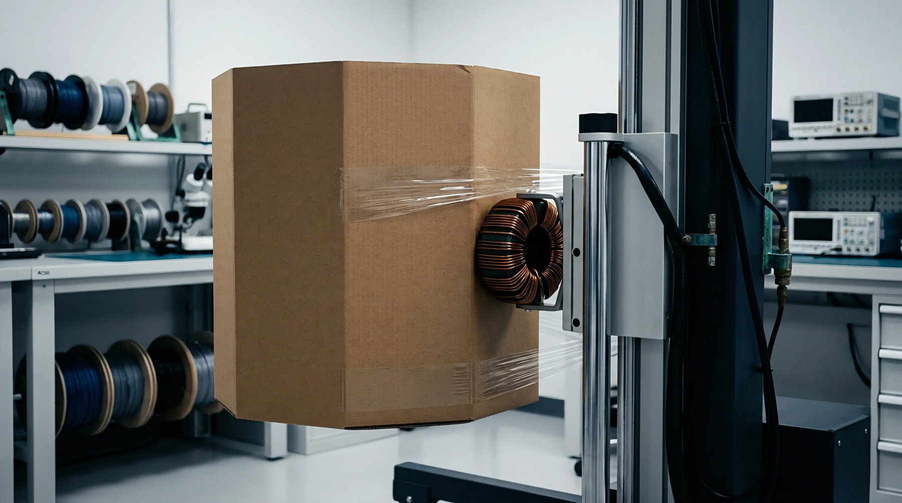
Automated scanning procedure captures the internal field distribution of a high-frequency source at a high resolution across a three-dimensional block of space. Performing a volumetric zoom scan identifies the exact spatial peak of radio energy inside a tissue-simulant phantom. This technique refines the initial rough area scan to find precisely where energy is concentrated to an infinitesimal point.
It contributes to the final assessment of the mass-averaged energy absorption during safety qualification.
Spatial Resolution
Precision mechanical arms move the measurement probe in small increments along three axes inside the grid. Every coordinate in the volumetric zoom scan grid yields a local field strength value used for interpolation later. High density data points ensure that even very small hotspots are correctly identified and measured.
This level of detail is necessary to distinguish between nearby signal sources on a single board.
Data Capture
Software analyzes the raw field readings to determine where the values begin to drop off rapidly. A volumetric zoom scan must fully capture the decline from the peak to confirm that the entire high-intensity region has been assessed. Typical grids consist of five to eight cubic centimeters depending on the frequency of the source.
The result is a numerical average that stays below the mandatory limit for human tissue exposure.
Peak Identification
Locating the highest intensity ensures that conservative safety margins are built into the host product design. Results from the volumetric zoom scan show exactly which part of the chassis generates the highest field leakage. If the scan area is too small, there is a risk that the actual peak value remains undetected.
Final dossiers include detailed graphical slices from these scans to show internal field contours.